Parallel beam X-ray optics are optical components with a graded multilayer deposited on a substrate having a parabolic shape in beam direction. These optical devices convert (in one dimension) a divergent incoming beam into a parallel one, or vice-versa an incoming parallel beam into a focusing one.
In order to obtain high efficiency, the d-spacing of the multilayer has to be varied from the front end to the rear end of the optics in correspondence to the aspheric curvature. Either the X-ray source or the detector (or detector slit) may be placed at the optics focal distance, for primary or secondary side applications, respectively.
X-ray Optics

Flat and curved multilayer X-ray optics can be used as monochromators, collimators or focussing optics in X-ray diffraction, X-ray reflectometry, X-ray fluorescence analysis and for synchrotron applications. Several types of multilayer X-ray optics can be designed depending on the customers application.
Parallel Beam X-ray Optics
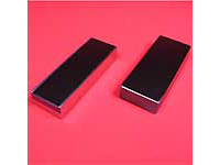
High precision parallel beam optics on bended wafer piece (left) and on prefigured substrate (right).
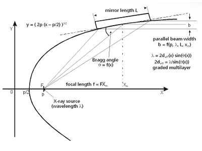
Parallel beam geometry in one dimension
|
Spectral lines |
Cr, Co, Cu, Ga, Mo, Rh, Ag, In, W |
|
Mean Reflectivity |
R > 70% |
|
Monochromaticity |
Kα1+Kα2 or Kβ |
|
Divergence |
Δφ < 0.03° |
|
Mirror length |
L = 40...100 mm |
|
X-ray source type |
line focus |
|
Parallel beam width b |
dependent on mirror length, geometry and X-ray wavelength, typical values: |
|
Typical focal length |
xm = 100..110 mm |
Focusing X-ray Optics
Focusing X-ray optics convert a divergent incoming beam into a focusing one. Having an elliptical figure of curvature the d-spacing of the multilayer coating has to be changed from the front end to the rear end of the optics. The primary and secondary foci of the mirror are located in the two focal points of the ellipse which typically coincide with the X-ray source point and the sample or detector position.

Parallel beam and focusing X-ray optics with various geometries
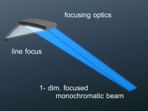
Generation of a monochromatic focusing beam in one dimension
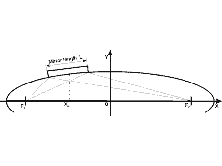
Beam path and main parameters of focusing X-ray optics
|
Spectral lines |
Cr, Co, Cu, Ga, Mo, Rh, Ag, In, W |
|
Mean Reflectivity |
R > 70% |
|
Mirror length |
L = 40 mm ... 100 mm |
|
X-ray source type |
line focus |
|
Focal line width b |
depending on spectral line, geometry and mirror length |
|
Typical focal lengths |
f1/2 = 100..2500 mm |
ASTIX-c: 2-dimensionally collimating
ASTIX-c mirrors are 2-dimensionally collimating X-ray optics in a modified Montel geometry (1) for the generation of 2-dimensional high intensity parallel X-ray beams.
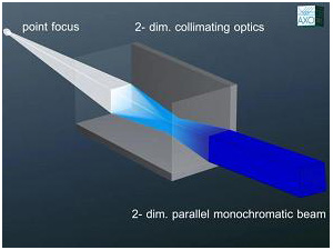
Working principle of ASTIX-c collimating geometry
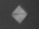
Parallel beam profile for Mo Kα radiation, I>107 cps (low power µ-source), b² ≈ 1 mm²
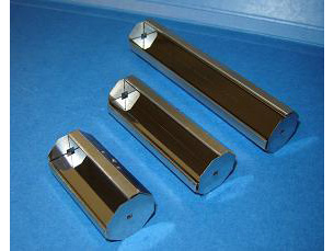
Photo of different ASTIX mirrors.
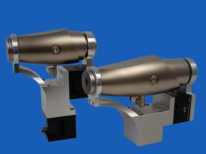
High precision vacuum mirror housings for ASTIX optics
|
Spectral lines |
Cr, Co, Cu, Ga, Mo, Rh, Ag, In, W |
|
Typical lengths |
60 mm - 150 mm |
|
X-ray source type |
all typical types of X-ray sources (rotating and fixed anodes, liquid metal jet and micro focus X-ray tubes) |
|
Typical parallel beam size |
1 mm² ≤ b² ≤ 5 mm² |
|
Typical focal length |
f1 = 100..110 mm |
|
Accessories |
High precision vacuum mirror alignment housing |
ASTIX-f: 2-dimensionally focusing
ASTIX-f mirrors are 2-dimensionally focusing X-ray optics in a modified Montel geometry (1) for the generation of high intensity focused X-ray beams. They can be optimized either for high flux or high resolution.
High Flux (HF) Optics
- high photon flux at sample position
- high integrated pixel intensity
High Resolution (HR) Optics
- small spot size at sample position
- high lateral resolution in the detector plane
(1) M. Montel - "The X-Ray Microscope with Catamegonic Roof-Shaped Objective" in: X-ray Microscopy and Microradiography, Vol.5, 1957,pp 177 - 185
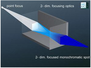
ASTIX-f: focusing geometry
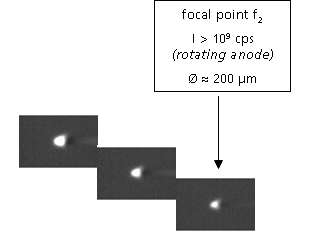
Profile of the diffracted beam between optics and focal point (f2 = 310 mm)
|
Spectral lines |
Cr, Co, Cu, Ga, Mo, Rh, Ag, In, W |
|
Typical lengths |
60 mm - 150 mm |
|
X-ray source type |
all typical types of X-ray sources (rotating and fixed anodes, liquid metal jet and micro focus X-ray tubes) |
|
Typical focal spot sizes |
<30 µm .. 500 µm |
|
Typical focal lengths |
f1/2 = 100..500 mm |
|
Accessories |
High precision vacuum mirror alignment housing |
Flat Graded X-ray Optics
Flat graded X-ray optics are mirrors with an artificial multilayer coating showing a lateral thickness gradient along the substrate length. They monochromatize the incoming beam while leaving the divergence unchanged, i. e. they generate a divergent monochromatic beam. In order to obtain high efficiency, the d-spacing of the multilayer has to be changed from the front end to the rear end of the mirror. Flat graded optics require a plane substrate and have a single focal point (line). Either the X-ray source or the detector (or detector slit) may be placed at the optics focal distance, for primary or secondary side applications, respectively.
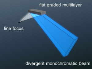
Generation of a monochromatic 1-dimensional divergent beam
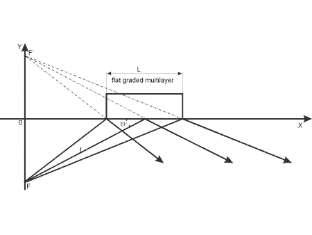
Beam path and main parameters of a flat graded multilayer
|
Spectral lines |
Cr, Co, Cu, Ga, Mo, Rh, Ag, In, W |
|
Mean Reflectivity |
R > 70% |
|
Monochromacy |
Kα1+Kα2 |
|
Mirror length |
L = 20 mm ... 80 mm |
|
X-ray source geometry |
line focus preferred |
|
Capture angle |
φ=0.20° (L = 20 mm) ... |
|
Application |
monochromator (primary or secondary) |
Monochromators and synchrotron mirrors
Monochromators are optical devices with a multilayer of constant period thickness over the substrate length in order to obtain high efficiency. A plane figure is usually required for such monochromators. Depending on the application either high resolution or high flux multilayer monochromators can be fabricated.
Download: Synchrotron optics prospectus (pdf)
Download: Tender X-ray optics prospectus (pdf)
|
Spectral range |
<50 eV - 100 keV |
|
Material systems |
optimized on wavelength or on customer's request |
|
Typical sizes |
up to 500 mm length (depending on parameters) |
|
Resolution |
0.25% < ΔE/E < 3% (periodic multilayers) |
|
Thickness homogeneity |
Δd/d < 0.2% |
|
Applications |
monochromators for laboratory X-ray sources and for synchrotrons, optimized for high reflectivity or tailored resolution polarizers in the soft X-ray range (O-K, Fe-L, Ni-L) |
Broadband Mirrors / Depth-Graded Multilayers
- Tailored multilayers with large number of different bilayer thicknesses to fulfill Bragg’s law for many photon energies / wavelengths
- Broadband or bandpass reflectors possible
- Energy bandwidths of 20% and more feasible
- Can be optimized for photon energies from EUV up to hard X-rays (80 keV and more)
- Selected bandwidth is maintained even if energy and corresponding incidence angle are changed
- Adaptation of the energy band to the source spectrum possible
- Large photon flux due to the collection of a large portion of the source spectrum e.g. at bending magnets
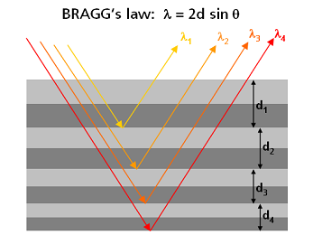
Working principle of a depth-graded multilayer: Different bilayer thicknesses reflect different photon wavelengths.
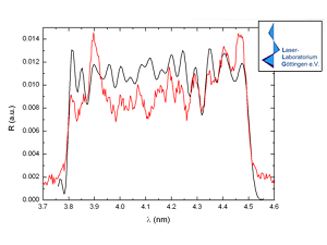
Reflected wavelength spectrum for a 17% bandwidth mirror around 4.13 nm (300 eV) at 45° incidence angle for polarization measurements with a laser plasma source (red: measurement, black: simulation)
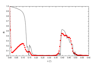
Reflectivity vs. grazing angle for a 22% bandwidth multilayer mirror at 22.2 keV (red: measurement, black: simulation)
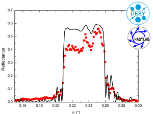
Reflectivity vs. grazing angle for a 21% bandwidth synchrotron mirror around 40 keV (red: measurement, black: simulation)